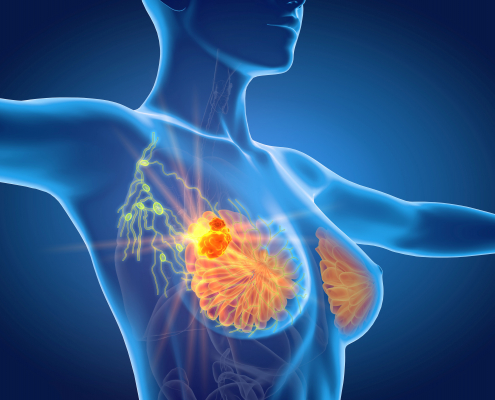
Contrast Enhanced Mammography (CEM)
Contrast Enhanced Mammography (CEM) is a special mammogram that uses iodine dye injected into the arm vein for better imaging. This dye makes it easier to find some breast cancers that may not be visible on a standard mammogram or ultrasound.
WHY DO I NEED A Contrast Enhanced Mammography (CEDM)?
Your doctor may recommend you have Contrast Enhanced Mammography for:
- evaluating a breast lump
- breast cancer screening if you are at increased risk, or have dense breasts
- breast cancer follow-up
CEM is a safe procedure. CEDM patients receive slightly more radiation than a regular mammogram, but it’s still within the recommended dose.
Some patients may have a reaction to the contrast dye. This is uncommon and usually very mild, such as itchiness or hives. Rarely, a reaction can be severe, such as shortness of breath or facial swelling. Our staff undergoes special training to treat these symptoms if they occur.
BreastScreen Victoria offers a separate breast screening service to Lake Imaging.
GEELONG BREAST CLINIC SERVICES
WHAT IS CONTRAST ENHANCED MAMMOGRAPHY (CEDM)
A contrast enhanced mammogram is a special type of mammogram that uses contrast dye containing iodine (similar to a CT scan) injected into an arm vein. This dye makes it easier to find some breast cancers that may not be visible on a standard mammogram or ultrasound.
WHY DO I NEED A Contrast Enhanced Mammography (CEDM)?
Your doctor may recommend you have Contrast Enhanced Mammography for:
- evaluating a breast lump
- breast cancer screening if you are at increased risk, or have dense breasts
- breast cancer follow-up
HOW DO I PREPARE FOR CEDM?
There is no special preparation required for Contrast Enhanced Mammography (CEDM). It is best to keep well hydrated beforehand and only eat lightly because some patients may feel mild nausea after the contrast dye.
Please inform our booking staff if you have kidney problems or are diabetic.
You may be more comfortable wearing a 2-piece outfit because you will be asked to remove clothing from the waist up and wear a gown.
WHAT HAPPENS ON THE DAY?
Your examination will be performed by a female technologist who has specialised in mammography. She will explain the procedure to you, and may ask you some questions about your symptoms and past history.
You will be asked to complete a questionnaire and sign a consent form. Our technologist will then place a small tube in the arm of your vein so that contrast dye can be given.
During the mammogram, each breast is briefly compressed for a few seconds. Whilst some patients find this uncomfortable, it should not be painful. Our technologists are specially trained to ensure this is as comfortable as possible.
As the contrast dye is given, you may feel a warm sensation which spreads through the body and a metallic taste in the mouth. This is normal. Please let the technologist know if you have any pain in your arm or feel unwell.
During the 3D component, you may notice the x-ray arm sweep in an arc, taking multiple low-dose images in a few seconds.
Most Contrast Enhanced Mammography take 15 minutes to complete. You may be asked to wait briefly whilst your images are checked.
AFTER A Contrast Enhanced Mammography (CEDM)
You will be asked to wait briefly whilst your images are checked. Sometimes further mammogram images are required, or an ultrasound is then performed. This is not uncommon.
If you have had no reaction to the dye, the tube in your arm will be removed, and a bandaid appliced. You can remove this when you get home.
It is important to drink 6-8 glasses of water after Contrast Enhanced Mammography to help clear the contrast dye from your kidneys.
WHEN DO I GET MY RESULTS?
Your Contrast Enhanced Mammography will be reported by a specialist breast radiologist, which can take some time because there are many images to look at and compare. Results are therefore not usually available immediately. We will send a report to your referring doctor.
Please ensure you have an appointment with your doctor to discuss these results.
PATIENT SAFETY
CEM is a safe procedure. Patients who have CEDM are exposed to slightly more radiation than a normal mammogram, but this is still within official dose recommendations.
Some patients may have a reaction to the contrast dye. This is uncommon and usually very mild, such as itchiness or hives. Rarely, a reaction can be severe, such as shortness of breath or facial swelling. Our staff are specially trained to treat these symptoms if they occur.
3D Mammography
(Tomosynthesis)
WHAT IS 3D Mammography (Tomosynthesis)
3D Mammography (Tomosynthesis) is new breast imaging technology that has been proven to improve the accuracy in diagnosis of breast cancer. It is performed as part of a diagnostic mammogram examination. In addition to the standard mammographic views, a specialised view (tomographic) is taken to produce 3-D images. Tomographic view is completed within seconds from a single sweep of the x-ray arm, creating a series of detailed images which together make up the 3D imaging of the breast.
For the tomographic view, the x-ray arm will move in an arc above the breast for a short time. Some women may experience discomfort with compression; however, if you experience pain during the mammogram you should inform the radiographer. You can also ask for the procedure to stop at any time.
BEFORE 3D Mammography
We recommend that you advise the radiographer if you have sensitive breasts, they will work with you to make sure that the mammogram is as comfortable as possible. Unfortunately, compression of the breast is essential to ensure an accurate image and minimise the amount of radiation used.
AFTER 3D Mammography
After the routine views of your breast have been obtained the radiographer will ask you to wait while they are examined by a radiologist to ensure that all the images needed have been obtained.
A breast ultrasound is often requested as a complementary test at the time of the mammogram or at a later date as a means of gathering more information for a complete examination.
Breast Hookwire
Localisation
What is BREAST HOOKWIRE LOCALISATION?
Mammography, ultrasound, and magnetic resonance imaging (MRI) examinations sometimes identify abnormalities in the breast that cannot be felt by a doctor.
If the abnormality is to be surgically removed, it is necessary to place a fine wire (called a hookwire), into the breast with its tip at the site of the abnormality. The wire acts as a marker during surgery and enables the surgeon to identify the correct area of breast tissue.
Mammography, ultrasound, or MRI scans are used to guide the hookwire into the correct position. The wire is called a hookwire because there is a tiny hook at the end, which keeps it in position.
BEFORE A BREAST HOOKWIRE LOCALISATION
A doctor’s referral and an appointment are required for this examination.
Please also bring along your request form, any previous imaging, and your Medicare card/any concession cards to your appointment.
Usually, this procedure will be performed a few hours before you have surgery. There is no preparation required for the hookwire localisation, but there will be preparation for the surgery that follows the hookwire localisation. Preparation instructions/information for the surgery will provided to you by your surgeon.
DURING A BREAST HOOKWIRE LOCALISATION
Before the procedure you be asked to remove all jewellery and clothing from the waist up and change into a loose-fitting examination gown.
The skin of the breast will then be washed with antiseptic before a very fine needle is used to give local anaesthetic to numb the breast in the area for biopsy. The local anaesthetic may sting for a few seconds when it is being given, and after this the area will become numb.
The radiologist will then insert a fine needle into the tissue to be removed. Images will be taken to check the position of the needle, once it is in the correct position, a fine wire is passed through the centre of the needle and the needle is removed, leaving the hookwire in place. A final set of images will be taken to show the surgeon where the tip of the wire lies in relation to the abnormality that is to be removed.
AFTER A BREAST HOOKWIRE LOCALISATION
Following the hookwire placement, a piece of the fine wire will be protruding from the breast. This projecting wire will be taped down to the skin and the hookwire remains in the abnormality in the breast. The surgeon will remove the wire together with the abnormality at the time of the operation. Your previous imaging and the images from the Hookwire Localisation will be sent with you to the operating theatre so that the surgeon may refer to them.
The purpose of the hookwire procedure is to provide a physical guide for the work of the surgeon. As it is not an investigation, there are usually no results for the hookwire procedure itself other than a written description of what was done and provision of the guidance images.
After surgery, the surgeon will give you the pathology result for the tissue removed, when you have your appointment with the surgeon after the operation.
PATIENT SAFETY
Hookwire localisation is a simple procedure to perform, and most women will experience no problems. Problems that can occur on rare occasions are;
• movement of the hookwire after placement and before surgery is performed (which reduces the accuracy of the surgery), and
• Wire dislodgement. This occurs usually because the breast is composed of fatty tissue which provides a poor grip for the hookwire).If you are travelling to another facility for your surgery with a hookwire in position, you need to take care. Dislodgement may occasionally occur with very little movement. If dislodgement occurs, you may need to have the procedure repeated because the tip of the wire will no longer be situated in the lesion that needs to be removed.
Breast Vacuum-Assisted
Stereotactic Core Biopsy
What is BREAST VACUUM-ASSISTED STEREOTACTIC CORE BIOPSY?
A Vacuum-assisted stereotactic core biopsy (VAB) is a biopsy which removes a sample of tissue from the breast for examination by a pathologist. The VAB is performed under mammographic guidance, allowing the area for biopsy to be accurately identified, using a special needle with suction to help get the sample from the breast.
A Vacuum-assisted stereotactic core biopsy is performed to help determine the nature or diagnosis of a breast abnormality and to plan treatment if necessary.
BEFORE AN BREAST VACUUM-ASSISTED STEREOTACTIC CORE BIOPSY
A doctor’s referral and an appointment are required for a breast ultrasound guided core biopsy.
Please let us know when making your appointment if you are taking medication that makes you bruise or bleed easily.
DURING A BREAST VACUUM-ASSISTED STEREOTACTIC CORE BIOPSY
When you arrive at Lake Imaging you will be asked to remove all jewellery and clothing from the waist up, and change into a loose-fitting examination gown.
VAB is performed while sitting or lying in a special chair with the breast compressed in the same way as for a mammogram. Several keyhole images will be taken to accurately identify the area for the biopsy.
The skin of the breast is then washed with antiseptic before a very fine needle is used to give local anaesthetic to numb the breast in the area for biopsy. The local anaesthetic may sting for a few seconds when it is being given, and then the area will become numb. A small incision will be made and several samples taken. The biopsy procedure may sometimes feel uncomfortable but is usually not painful because of the local anaesthetic that has been given. The process of taking the biopsy lasts just a few minutes.
After the samples have been taken, the biopsy area will be held firmly for a few minutes to minimize any bruising or bleeding, and then covered with a dressing that will be checked before you leave.
Please allow about an hour for the procedure.
AFTER A BREAST VACUUM-ASSISTED STEREOTACTIC CORE BIOPSY
After the procedure, the biopsy site may be tender or show some bruising. You may place an ice pack over the biopsy site for no more than 20 minutes. We suggest taking Paracetamol for discomfort. You will be able to leave the clinic shortly after the procedure.
• You may drive yourself home after the procedure.
• Most people can return to work the same day.
• You should refrain from exercise for 24 hours following your biopsy.
• Your dressing may be worn in the shower and removed after two to three days.If your breast becomes red, swollen or tender in the days after your biopsy please consult your doctor or contact the Lake Imaging clinic to review the biopsy site.
Results from VAB are usually available within 48 hours, but may take up to a week. Our nurses will ensure that you have an appointment either in our results clinic or with your referring doctor to get your results as soon as possible.
PATIENT SAFETY
VAB is a safe procedure, and most people do not find it uncomfortable. Please advise the radiologist if you are experiencing any pain.
After the local anaesthetic wears off, you may experience some discomfort in the breast, but this is usually eased by taking paracetamol.
Fainting during the biopsy is uncommon because you will be sitting or lying during the procedure, but please let the doctor know if you are prone to fainting.
Most people develop some bruising of the skin at the biopsy site lasting for a few days after the procedure. Sometimes a bruise may develop within the breast tissue which may cause a tender lump. This may take one to two weeks to disappear.
Infection can sometimes occur when tissue is cut, but this is rare following VAB. If your breast becomes red, swollen or tender in the days after your biopsy, please consult your referring doctor or contact Lake Imaging to review your biopsy site.


